
Docs /Medical Workflow Segment and Reconstruct
Segmentation Methods
Segmentation is the process of identifying and isolating anatomical structures in medical images. In LeaRes, segmentation and 3D reconstruction live in a single workflow opened from the Segment and Reconstruct panel.
Segmentation Overview
Why Segment?
Segmentation enables:
- 3D Visualization: Create surface models of organs
- Functional Analysis: Run virtual spirometry and surgical planning on lung anatomy
- Simulation: Turn an anatomy into a CFD project (the bridge to the engineering workflow)
Segmentation Methods
The panel offers three methods, selected from the method selector at the top:
- Auto segmentation (default): server-side AI on GPU. Organ groups come from pre-trained SegResNet checkpoints exported from MONAI Auto3DSeg (one each for cardiac, lungs and whole body). Two optional add-on models run as background jobs: TfeNet for airways and nnU-Net for coronary arteries.
- Region growing: seed-based growth controlled by an intensity threshold
- Thresholding: select voxels within an intensity range
Note: Voxel segmentation is driven by the three methods above. Two interactive tools sit outside this panel: Scissors (toolbar, 3D image carving) draws a spline contour on a slice and cuts the volume inside or outside it, with undo/redo; and Edit mask (Geometry editing group) offers a sphere/cube brush for adding or removing regions. Edit mask is not yet wired to the backend in this build.
Opening the Segmentation Panel
- Load a DICOM acquisition in the viewer (see Uploading DICOM Data)
- In the vertical toolbar, click Segment and Reconstruct
- The panel opens at the top of the left Data sidebar, above the tree
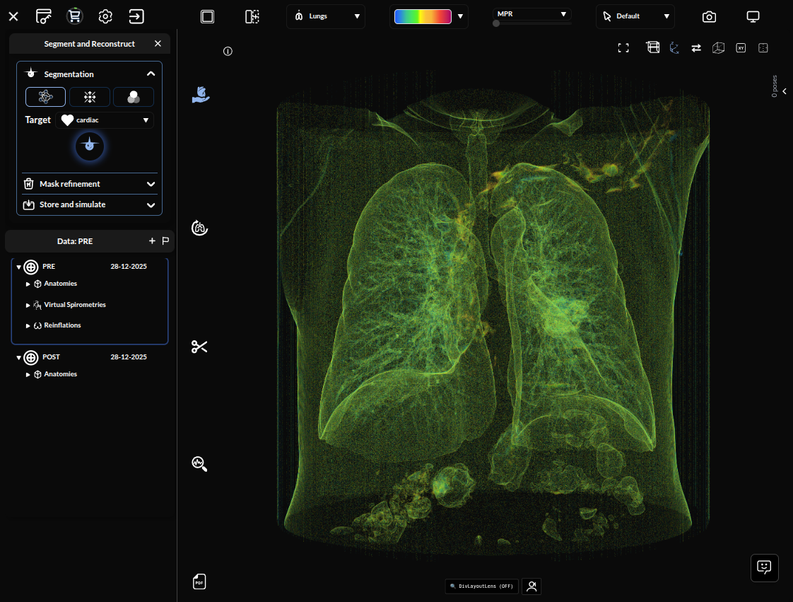
The panel contains:
- Method selector: Auto segmentation, Region growing, or Thresholding
- Method controls: parameters for the selected method
- Select segments: the list of produced segments
- Surface post-processing lives in the Geometry editing group on the vertical toolbar (covered in 3D Reconstruction)
- Store selection as project: save the anatomy as a project/device
Method 1: Auto Segmentation
Auto segmentation uses AI models running on the server (GPU) to identify anatomy automatically.
Running Auto Segmentation
- Make sure Auto segmentation is selected (it is the default)
- Choose the anatomy to segment from the Target dropdown (default
cardiac) - For the lungs, optionally enable the Segment airways (TfeNet) checkbox to also extract the trachea and bronchial tree
- Press the round Start button
The job runs server-side on GPU. When it finishes, the produced segments appear in the Select segments list and an initial 3D surface is generated automatically.
Tip: Enable Segment airways (TfeNet) whenever you plan to run virtual spirometry — airway geometry improves the downstream lung analysis.
Method 2: Region Growing
Region growing segments a connected region that shares similar intensity values, starting from a seed you place on the volume.
Using Region Growing
- Select Region growing in the method selector
- Click Add Seed, then Alt+Click inside the target structure in any 2D view (axial, sagittal or coronal). Press Esc to remove the seed.
- Adjust the Threshold range — it opens centred on the seed’s HU value ±100, within a ±300 track
- Set Smoothness (1–10, default 4) to control how much the resulting surface is smoothed
- Press the run button, or Enter, to launch region growing
- Press Backspace to discard the resulting Region growing mask and start over
Tip: Place the seed away from organ boundaries and large vessels to avoid the region leaking into neighbouring tissue.
Method 3: Thresholding
Thresholding selects every voxel whose intensity falls inside a chosen range, regardless of connectivity.
Using Thresholding
- Select Thresholding in the method selector
- Set the Threshold range — it defaults to −500 … +500 HU on a −1000 … +1000 track — and the Smoothness level (1–10, default 4)
- Press the generate button to launch the segmentation; the voxels inside the range form the segment
Approximate CT reference ranges (Hounsfield Units):
| Tissue | HU range |
|---|---|
| Air / lung | -1000 to -700 |
| Fat | -100 to -50 |
| Water | ~0 |
| Soft tissue | 20 to 100 |
| Bone | 200+ |